Book review and guest post – ‘The Little Book of Honey’ by Elizabeth Gowing

When I first started writing this blog I thought it would pretty much just be me reading it. I never imagined that one day a writer I greatly admire would ask me to review their book! Yet this day has arrived, and the lovely Elizabeth Gowing, author of ‘Travels in Blood and Honey: Becoming a beekeeper in Kosovo‘, has sent me her new book, ‘The Little Book of Honey‘.

Little Book of Honey cover In the first part of the book, Elizabeth gives us her opinions of honeys she’s enjoyed from around the world. It is a fascinating way of tasting your way around different parts of the Earth, taking in the concentrated scents and tastes of the flowers surrounding the bees. Is there any other food that reflects its local area so vividly?

Elizabeth’s lushly descriptive writing brings the labours of the bees to life. She even manages to find a honey “better than chocolate” – carob honey. And viper’s bugloss reveals itself as having a fairy tale taste, “with a butterscotch and ripe banana taste”.

The second half of the book contains honey recipes, some of which come with particular honeys recommended. Apple blossom honey is the best to have with a croissant. French honeysuckle is ideal for making ice lollies and Greek pine honey for Loukoumades, a Greek dessert of fried dough balls soaked in honey syrup.

Naturally I couldn’t review the book properly without first trying out its recipes. Drew and my colleagues have generously helped me out with tasting the resulting cakes. The first recipe I tried was Honey Spice Cake, which fills the house with the sweet smells of cinnamon, ginger, allspice and cloves. This went down well, the honey taste really coming through.  My mum’s family is Welsh, so for my second recipe I chose the Welsh Honey Muffins. I like fruity cakes, so I added in some sultanas soaked in honey flavoured tea, and also made them fairy cake size. Dark and full of flavour, yet very soft and airy in texture, they brought the uplifting scents of cinnamon and ginger amidst the darkness of winter.

Honey muffins

Honey muffins

I have a couple of minor niggles with The Little Book of Honey as a recipe book – size and measurements. It’s small size is sweet but means the book needs weighting down to keep it on the right page whilst baking. And although it’s published in the UK, the measurements are in US cups (though there is a conversion chart at the back). My problem with US cups is that I don’t feel the measurements are as accurate, especially when it comes to measuring butter. Slabs of butter don’t fit properly into a round cup. Anyway, these are tiny complaints – the vivid descriptions and unusual recipes more than make up for them.

I asked Elizabeth if she could explain a little about the background of the book, how she came to write it and went about researching it. This is what she said:

“I’ve always been keen on buying local and when I was travelling around, and particularly when I came from England to live in Kosovo, I discovered that honeys from different landscapes and flowers have a ‘terroir’ just like wine, and you can taste the combination of flowers whose nectar contributed to any particular jar. Just like wine, every batch of honey is unique too and I got excited by tasting honey from coriander flowers, from avocado flowers, pine honey in Greece, ivy honey in Cornwall (strangely like Stilton), beechwood honey from New Zealand etc etc.

When I became a beekeeper I also became interested in what to do with the honey I harvested (other than just spreading it on toast!) and researched some recipes and realised there were lots more things I could do with the honey – smoothies and breads, desserts and snacks. I’ve included in the book recipes for Russian ‘sbiten’ herbal honey drink, Yemeni honey bread, Welsh honey muffins (my favourite recipe in the book), and honey vinaigrette.

Many of the recipes I found came from other beekeepers and in talking to them I also heard about some ways that beekeeping can change lives – a poverty reduction project in Nigeria with Bees Abroad, the inspirational work of the Golden Company with disaffected young people trained as beekeepers in Hackney, or the women in a shelter in Kosovo who have survived trafficking or domestic violence and who now have a business producing wonderful face creams with the propolis from their beehives to support themselves. When you hear stories or savour tastes like these you want to share them, so I had the idea of the Little Book of Honey.

I’d worked with designers, Su Jones and Paddy McEntaggart on the design for my first book, Travels in Blood and Honey; becoming a beekeeper in Kosovo (Signal Books, 2011) so I knew they would transform my words into a beautiful artefact. I think what they’ve produced is really gorgeous – with woodcut-style illustrations on every page it’s a treat to hold and flick through (check out the flickbook effect of the bees around the page numbers!) and the perfect gift for any beekeeper or foodie. People can see some sample pages – and order a copy! – at the website www.thelittlebookofhoney.co.uk. The book costs £6 plus £1.50 P&P.”

Order a giftpack with a signed copy of my Travels in Blood and Honey; becoming a beekeeper in Kosovo, together with a hand-crafted Kosovan honey drizzler. See more at http://100daysofhoney.wordpress.com/2011/11/12/a-new-way-to-eat-honey-my-own-kosovan-handcrafted-drizzler-also-available-to-buy/

Fascinating – thanks so much Elizabeth! I love my Little Book of Honey; it has a place of honour on my bee themed bookshelf.

Related posts/links:

Posted in Uncategorized | Tagged | 18 Comments

Beekeepers in the snow

It takes a certain type of lunatic person to be a beekeeper. That type of person is prepared to turn up and talk bees, drink tea and eat cake in all weathers, and that includes snow!

Ealing beekeepers in the snow

From left to right: David, Don, Albert, Stan, John and Cliff.

A great selection of hats going on there.

A small amount of beekeeping was also done. This consisted of hefting hives, checking fondant levels were still ok, and looking at a woodpecker hole that had appeared in the bottom of John’s hive. Worrying, as once a woodpecker has learned the habit of drilling into beehives they are likely to try it again. To a hungry woodpecker, our apiary must be like a piñata waiting to happen. John covered over the hole with parcel tape.

Snowtracks III

Are these the claw prints of the culprit? Or perhaps they belong to a seagull or wood pigeon?

Snow tracks

And could these smaller paw prints be those of a squirrel?

Snowtracks II

Naturalist David Craven posted a great idea on his ‘Why watch wildlife‘ blog – taking photos of the prints we find in the snow, and then tweeting them with the #snowtracks hash tag. It’s not often we get to see what creatures have been visiting! Have you come across any snow tracks for David to identify?

Emma photographing the bees

Emma photographing the bees

We peeked inside our hive and the bees were still busy eating through their fondant. Emma took a close-up photo of them – you can see the result on her latest blog post, ‘Snowmageddon‘.

What’s the weather like in your area, are your bees under snow or enjoying sunshine?

Posted in Uncategorized | Tagged | 22 Comments

9th Honey bee pests, diseases and poisoning revision post: chilled brood and neglected drone brood

A 9th revision post for the British Beekeeping Association’s Module 3 Honey bee Pests, Diseases and Poisoning exam, which I’m taking in March. I’m doing the BBKA’s correspondence course, so my tutor, Pam Hunter, sets me papers which she then marks. The second question of Paper 3 is all about chilling, very appropriate as it’s mighty bitter out there just now…

B2. a) discuss the possible causes of chilled brood

  • A lack of bees

Any factor which reduces the number of bees in a colony below the minimum needed to look after the brood will cause chilling; this could include poisoning, loss of a large swarm, a rapidly growing colony during spring or plain old beekeeper cock-ups.  The effects can be seen when large areas of brood fail to develop, whereas disease usually causes a pepperpot effect with some larvae emerging and others failing.

Photo of chilled brood.

Photo of chilled brood. Courtesy The Food and Environment Research Agency (Fera), Crown Copyright

The bottom and sides of brood frames are most likely to contain chilled brood, and it is most likely to be seen in early spring, when the temperature may drop after a warm spell which encouraged the queen to lay well. Dead brood can first appear a yellowy grey colour, before turning dark brown or black, then finally drying out to removable black remains. Nurse bees will abandon the frames on the outside of the brood nest first and try to preserve the larvae in the warmer centre.

  • Beekeeper mishandling

Don’t try to make up nucleuses or hives with an imbalance in the ratio of brood to adult bees; make sure there are plenty of bees to look after the brood. The BBKA has produced Nucleus guidelines which recommend that “Brood and eggs in all stages should occupy at least half the total comb area, with no brood cycle break…There should be a good balance of young and flying bees, and the frames should be well covered.”

A well-meaning beekeeper trying to speed up the colony’s growth by ‘spreading’ or moving about the brood frames by putting extra frames of foundation between brood frames can cause chilling. Do not be tempted to mess with the brood nest!

Long inspections in chilly weather should be avoided. However, bear in mind that young larvae can survive for several hours at well below normal brood nest temperature (35C/95F). Chilled brood is caused by prolonged exposure to cold temperatures and/or starvation due to neglect by the adult and nurse bees in the colony – not beekeeper inspections. But leaving the brood box open for several minutes in cold weather isn’t going to help or endear you to the bees.

To help the bees in keeping the brood warm, try not to give them too much space whilst the colony is building up in spring, particularly if you are filling in the brood box with empty frames of foundation. If you have a small colony in a ten frame brood box but they are only on four frames, the sixth frame down can be replaced with a solid wooden ‘dummy board’, to help keep warmth in. The dummy board can then gradually be moved out as the colony occupies more frames.

b) short periods of chilling can make brood susceptible to another brood disease; name the disease and its causative organism

Chalkbrood is an extremely common brood disease which is often present at low levels in colonies. It is thought to become a noticeable problem when the colony is weak and when levels of carbon dioxide rise above normal, because the bees are failing to maintain the correct conditions in the hive. It is also linked to stresses such as insufficient nurse bees, pollen shortage and the presence of sac brood.

Chalk brood infection

Chalk brood infection. Courtesy The Food and Environment Research Agency (Fera), Crown Copyright.

Chalk brood is caused by a fungus named Ascosphaera apis. This delightful organism begins to germinate when a larva takes in its spores with its food. Inside the gut, the spores start to grow, producing multiple branches of fine cotton-like threads. These break through the gut wall and continue to grow throughout the body of the poor larva, until eventually it becomes “a swollen mass of fluffy white fungus with a small yellow lump where its head used to be“, as Celia Davis puts it in her excellent book ‘The Honey Bee Around & About’ (2007).

The infected larva dries to a hard chalk-like lump called a ‘mummy’, which can be white, grey or black. These will rattle when the comb is shaken. Death occurs after the cells have been sealed, so workers will tear the cappings open to remove the mummies and dispose of them outside the hive. Unfortunately the mummy spores are sticky and will attach to the bees, causing them to infect larvae when they re-enter the hive. Yet another reason to change brood comb regularly – the spores are resistant to heat and have a life of between 3-38 years.

Chalk brood mummies on a hive floor.

Chalk brood mummies on a hive floor. Courtesy The Food and Environment Research Agency (Fera), Crown Copyright.

Like chilled brood, beekeepers are most likely to see chalk brood in the spring when colonies are expanding the brood nest rapidly, but do not yet have a large adult bee population. Even if the resulting chilling is not sufficient to kill the larvae, it seems to encourage the growth of the Ascosphaera fungus.

c) what are the causes of neglected drone brood?

Drones are more dispensable than workers. They eat lots, do no work and generally slob about the hive. Their bulk can be useful in keeping the brood warm, but still they are less than essential for the individual colony to survive.

Drone face

Not particularly useful, but they are handsome. Photo taken by Drew Scott.

So drone brood is usually the first to be neglected and ignored if a colony is under pressure, for instance if little forage is available or long periods of bad weather stop the bees foraging. The sisters may then unceremoniously ditch their brothers to ensure the colony’s survival, often throwing the drone larvae out of the hive.

In the case of a drone-laying queen or laying workers, no new nurse bees are being produced, so the increasingly old workers are unable to keep up with feeding all the drone larvae.

References:

Posted in Disease prevention, Exams | Tagged | 16 Comments

8th Honey bee pests, diseases and poisoning revision post: collecting laboratory samples

An 8th revision post for the British Beekeeping Association’s Module 3 Honey bee Pests, Diseases and Poisoning exam, which I’m taking in March. I’m doing the BBKA’s correspondence course, so I have a tutor setting me papers which she then marks. I’ve just sent my answers to Paper 2 off to her, now on to Paper 3 and a look at collecting lab samples if you suspect disease or poisoning in your hives…

B1. a) what is the recommended sample size needed for the diagnosis of:

  • i) adult bee diseases

30 bees must be examined in order to complete the diagnosis for acarine, nosema and amoeba.

  • ii) poisoning by toxic chemicals

At least 200 dead bees are required for a full analysis for a poisoning sample, approximately the number that will fit into one of the large ‘Cooks’ matchboxes. The National Bee Unit advises that this needs “to be securely packaged, not in a plastic bag and preferably not flattened“.


Photo from the ‘How can I recycle this‘ website.

If possible, at the same time take two further samples of 200 bees and put these in the freezer in case they are needed. Note the time and day when the bees are discovered and try to locate any witnesses to spraying.

  • iii) why these minimum sizes?

With a sample size of 30 bees, there is a 95% probability that acarine, amoeba and nosema will be detected within this sample, if the infection level is serious enough to require treatment.

Some beekeepers believe there is a strong case to be made for shifting our assessment of nosema infection from “intensity” (as measured by spore counts) to “prevalence” (the percentage of bees actually infected).

Courtesy The Food and Environment Research Agency (Fera), Crown Copyright

Analysing a sample. Courtesy The Food and Environment Research Agency (Fera), Crown Copyright

b) how would you collect these samples?

You want the samples to be fresh but the bees to be old…

Choose bees that are the freshest, submitting newly dead or dying bees if possible.

For some diseases, such as nosema, it is best to collect older foragers. This is because older bees have higher spore counts, since nosema in the gut multiplies and increases during an infected bee’s life. As young bees under 8 days of age won’t have any spores, sampling them could lead to “false negatives.”

Martin-Hernandez (2006) found that foragers returning to the hive can display up to 10-fold higher infection levels than interior bees, and that both interior and exterior bees had almost double the spore count when sampled at 12:30 pm rather than 8:30 am (Scientificbeekeeping.com, the ‘“Nosema Twins” – Part 3, Sampling’ article).

As well as being at the hive entrance, inside the hive foragers are more likely to be at the edges, such as the lid and outside combs. An easy way to gather these bees is to scoop them up from the inside surface of the crown board.

If bees are crawling in front of the hive entrance because they are too weak to fly, these are easy to collect and just as likely to contain nosema spores or viruses as foragers.

Lab training

Microscopy workshop. Courtesy The Food and Environment Research Agency (Fera), Crown Copyright


c) how would you kill a sample of bees for adult bee disease diagnosis at home?

Live bees can be killed by putting them in the freezer overnight.

d) how should the samples for laboratory confirmation be prepared and packed for sending by post?

  • Brood disease samples

The best method is for beekeepers to submit a whole brood comb, with brood of all stages present, in order for an accurate diagnosis to be made.

  • Adult bee disease samples

The best way to send in adult bee samples is in a small sturdy box, well packaged in something such as a padded envelope so the bees will not be squashed. A suitable size for the box is a standard-sized matchbox, which will contain 30 bees. Avoid using glass or plastic boxes, which will degrade the sample by hastening decomposition.

  • Poisoning samples

At least 200 bees are required for a full analysis for a poisoning sample, approximately the number that will fit into one of the large ‘Cooks’ matchboxes. The National Bee Unit advises that this needs “to be securely packaged, not in a plastic bag and preferably not flattened“.

  • General advice

Dry out any wet bees that have been exposed to the elements on newspaper before packing.

Indicate which diseases you want the bees examined for and include a contact phone number in case the lab has any questions about your sample. A template voluntary sample submission form can be downloaded from the National Bee Unit’s Beebase website and printed off. Information which might assist with the diagnosis should be given in a covering letter.

A cheque made payable to the Food and Environment Research Agency should be included; the 2012 price for lab analysis of a sample for Acarine, Nosema and Amoeba is £40.00, but check the National Bee Unit (NBU) prices page for current prices.

‘Bees’ written clearly in large letters on the outside of the parcel will often ensure swift delivery. You don’t have to tell the posties that the bees are dead!

e) to whom could these samples be sent?

Beekeepers in the UK can send samples to the National Bee Unit’s laboratory diagnostic team in York.

FERA NBU Beesuit

A NBU inspector’s bee suit. Courtesy The Food and Environment Research Agency (Fera), Crown Copyright

f) give a list of crops most likely to be sprayed with chemicals harmful to honeybees?

Farmers or gardeners are most likely to spray fruit trees, soft fruit and field crops such as oil seed rape, beans, borage, vegetables, legumes and cereals.

Local authorities may spray weeds such as poppy, ragwort, dandelion and charlock.

Have you ever had to submit a sample for lab analysis? How did you go about it, and what were the results?

References:

Posted in Disease prevention, Exams | Tagged | 14 Comments

Mysterious search terms used to find a beekeeping blog

Inspired by this amusing post on Steven Schwartzman’s ‘Portraits of Wildflowers’ photography blog, I thought I’d take a look at some of the more unusual search terms people used to find my blog in 2012.

‘woodlice droppings’ – 16 times

Urgh. I have failed to find any photos of woodlice droppings on my blog to keep these people happy. My internet research suggests that woodlice droppings are about 1mm in width and look like black dust particles.

‘what do woodlice look like’ – 4 times

Really? This is a bee blog! Oh well, if you insist:

woodlice photo
From www.uksafari.com.

do woodlice lay eggs – 1 time

Yes. A female woodlouse will keep fertlilised eggs on the underside of her body until they hatch into small, white offspring. That’s enough about woodlice now.

‘squashed bee’ – 12 times

Oh dear. I’m not very optimistic for this bee. Here’s one I’m afraid I accidentally squashed and is long since deceased.

Squashed bee dragging sting

ouch! that bee just stung me! – 2 times

Ouch indeed.

‘what do hives look like on adults’ – 6 times

Wrong type of hives.

‘what causes hives under the armpits’ – 2 times

Not bees.

‘hand feeding carrot’ – 3 times

Like this? Bulls love tasty crunchy carrots!

Bull eating carrots

Like this?

 ‘hunny from bees’ – 3 times

Mmm my favourite hunny.

Hunny

‘british beekeepers beware sign’ – 2 times

A sign to deter British beekeepers or a sign to warn others that British beekeepers may be present?

‘honey dripping on body’ – 2 times

Think they wanted a different kind of blog!

‘average monthly rainfall and temperature ealing’ – 2 times

In 2012, lots and flipping cold.

‘jon and sarah pie and mash wedding’ – 1 time

Huh?

‘bee pic taken by me ;)’ – 1 time

Try looking on your own computer? 😉 I did and here’s one I took earlier.

Honey bee on thistle

Honey bee on thistle

Posted in Uncategorized | 43 Comments

7th Honey bee pests, diseases and poisoning revision post: Being a bee detective – investigating the death of a colony

A 7th revision post for the British Beekeeping Association’s Module 3 Honey bee Pests, Diseases and Poisoning exam, which I’m taking in March. I’m doing the BBKA’s correspondence course, so I have a tutor setting me papers which she then marks. The last question for paper 2 is a very practical one – trying to work out what has caused a colony to die out.

B5. a colony was fed three gallons of syrup in late October during a period of rather damp weather; next Spring it was slow to build up compared with other colonies in the apiary; there were brown stains on the frames and combs; it recovered enough to produce swarm cells in early July but several of the queen cells had dark coloured walls and the occupants died in the propupal and pupal stages; on sunny days several workers could be found crawling outside the hive or clustered together on grass stalks with what appeared to be bloated abdomens; their wings looked somewhat dislocated and they were unable to fly.

a) give reasons to suggest what diseases might be present in this colony

  • Nosema

The information that the colony was fed three gallons of syrup during damp October weather and had brown stains on the frames and combs suggests the bees were suffering from dysentery.

Dysentery is not a disease but a symptom of problems in the bees’ excretory systems. It happens when excess water accumulates in the rectum, causing runny poop and increased brown splats around the hive’s entrance as the bees struggle to hold in. It is not a sign of nosema, but can help spread nosema spores if nosema is present.

dysentery stains

Defecation on a hive by bees with severe dysentery. From extension.org.

The suspicion that nosema is likely to be affecting the hive comes from the phrase ‘next Spring it was slow to build up’. This is the only observable symptom of nosema. The reason it stalls the progress of colonies is that the germinating nosema spores inside the gut of an infected bee impair the digestion of pollen, shortening the lifespan of the bee. Infection is particularly devastating in younger bees, which will not be able to digest enough pollen to develop their hypopharyngeal glands properly and be able to produce brood food.

  • Black queen cell virus

We may have had a brush with this in one of our colonies this year. Back in June, in my post ‘Long live Queen Neroli, our Jubilee Queen’ I recounted how we were worried at finding a queen cell still capped when the queen should have already emerged:

“We asked John Chapple’s advice. He instructed us to shake the bees off the frame and then gently cut the tip of the queen cell off with a hive tool to see what was inside. The queen revealed was obviously a goner, and had been for some time. “I’m no expert” (hah!) John said, “but that looks like black queen cell virus”. Having looked it up at home, this is a virus associated with the spore disease nosema, which is worrying. You can see her tattered wings and black body below.”

This queen, found in a queen cell from one of our hives in 2012, possibly died from Black queen virus. Copyright Emma Tennant.

This queen, found in a queen cell from one of our hives in 2012, possibly died from Black queen virus. Copyright Emma Tennant.

Black queen cell virus, which turns queen cell walls dark brown-black and the dead prepupa or pupa inside a yellow colour with a tough skin, is associated with nosema, which may be why the queen cells in the hive we’re investigating have succumbed. The virus can only multiply when nosema is present, and in this case several cells have dark coloured walls. The virus probably enters the developing queen through her gut, so as nosema weakens the gut wall this will allow the virus easier entry than the walls of a healthy gut would.

Our dead queen photographed above is black rather than yellow, so John may have been mistaken about the cause of death being Black queen cell virus.

Normally most honey bee viruses exist in a temporarily inactive state and do not cause signs of disease. However, the presence of other parasites or diseases can activate them. Viruses generally are often transmitted through diseases such as acarine, nosema and varroosis, which act as a vector by in some way facilitating the entry of a virus, or activating previously dormant viruses already present in the host. Varroa is thought to be a particularly common vector and viral replication has been shown to be activated by the presence of varroa. As the mites suck the haemolymph of the bees they also act as tiny hypodermic syringes, injecting the viruses directly into their hosts.

Varroa infestation - © Crown copyright 2010 "Courtesy The Food and Environment Research Agency (Fera), Crown Copyright"

Varroa infestation – © Crown copyright 2010 “Courtesy The Food and Environment Research Agency (Fera), Crown Copyright”

In ‘The Honey Bee Around & About’ by Celia F Davis (2009), she says:

“To a honey bee virus a bee hive must be paradise and when an organism such as Varroa destructor appears on the scene, to act as an efficient vector, it must seem like its Birthday and Christmas rolled into one.” (p39)

  • Chronic bee paralysis virus (CBPV)

The beekeeper has noted that “on sunny days several workers could be found crawling outside the hive or clustered together on grass stalks with what appeared to be bloated abdomens; their wings looked somewhat dislocated and they were unable to fly.”

Bloated, distended abdomens (due to a build-up of fluid in the honey stomach) and dislocated wings, causing inability to fly, with crawling on the ground or up plant stems, are symptoms of Chronic bee paralysis virus. There are two main ways the virus can manifest itself in colonies; one causes the problems noted above – abnormal trembling of the body and wings, bees crawling on the ground due to flight inability, bloated abdomens, and dislocated (K letter-shaped, partially spread) wings.

The other form causes hairless, shiny, and black-appearing bees that are attacked and rejected from returning to their colonies by entrance guard bees. The loss of hair is due to their hive mates having nibbled the hairs away. As they are denied entrance to their colony, their flight in front of the hive will be zig-zagging in a similar way to robber bees. These bees will also tremble.

Both forms of symptoms can be seen in bees from the same colony, and may reflect differences among individual bees in inherited susceptibility to the virus (from the Beebase website).

This photo shows three bees with typical CBPV

This photo shows three bees with typical CBPV “shiny appearance”. Courtesy The Food and Environment Research Agency (Fera), Crown Copyright.

During July the worker bee population would be at its peak. It seems that close contact amongst overcrowded bees can break hairs from the cuticle, allowing CBPV to spread from diseased bees to healthy bees via their exposed epidermal cytoplasm. It is likely that any factors that result in decreased foraging activities and crowded conditions in the bee colonies may lead to disease outbreaks of CBPV. It is also associated with varroa, and probably acarine.

b) how could the presence of the pathogens be confirmed?

Nosema

Definite confirmation of nosema requires microscopic examination of the abdomens of older adult bees, which can be collected from the hive entrance. Infection is highly variable in a colony, but foragers tend to contain the higher spore loads. Collecting bees leaving to go on cleansing flights rather than returning will be more effective. Send a sample of 30 bees to a microscopist or the UK National Bee Unit service in a small sturdy box (not plastic, which causes rapid sample degradation).

Lab investigation of a sample will also be needed to positively identify bee viruses.

c) how may the beekeeper have been partially responsible for the condition of the colony?

The information given tells us that the colony was fed three gallons of syrup in late October, during a period of rather damp weather. Sugar syrup feeding generally needs to be finished by early October in Britain, as if syrup is fed too late in the year the bees will have insufficient time to evaporate the excess water, and the syrup will be stored and could ferment, causing digestive problems for the bees when they come to feed on it later. Dysentery is caused by excess water accumulation in the rectum, due to diet or if the bees have been unable to fly for several days due to bad weather.

This late feeding of sugar syrup in unsuitable weather by the beekeeper probably caused the bees to develop dysentery and defecate within the hive, spreading nosema spores already present in the hive more quickly. The younger house bees will attempt to clean up the faeces, causing them to come into contact with the nosema spores in the faeces and become infected.

Widespread nosema infection could have weakened the colony, making it slow to build up in the spring and transmitting black queen cell virus to the queens in their blackened cells. This probably also left the colony more vulnerable to varroa and viruses associated with varroa, such as chronic bee paralysis virus. All this exacerbated by an unfortunate feeding choice in the autumn!

Dead bees in feeder

Sugar syrup in a contact feeder.

d) is there any advantage in uniting this colony to another one; give reasons?

The colony may currently be queenless if it has been unable to produce a healthy queen. If this is the case, uniting it with a queen-right colony will give it a chance of survival. As several workers have been observed unable to fly it is certainly likely to be low in numbers, and may be unable to build up enough stores and new bees to last overwinter without uniting.

There is possibly a genetic susceptibility to Chronic Bee Paralysis Virus, so requeening with a queen from a different strain is often recommended in bad cases. Uniting the afflicted colony to another one would fulfil this.

The disadvantage will be the chance that this colony could infect a healthy one. The beekeeper will have to weigh up whether it is worth the risk.

e) why may it be advantageous to fumigate the combs?

Fumigation would destroy the spores of nosema and several other infectious diseases which might be present on the combs. It is very difficult to treat honey bee viruses, so a good approach is to treat for vectors such as nosema, to try and reduce the exposure of bees to viruses.

Do you agree with this diagnosis? Have you ever come across similar symptoms in your hive?

References:

Posted in Disease prevention, Exams | Tagged , , | 18 Comments

A poem to mark the winter solstice

Today’s winter solstice, the shortest day of the year, has become important to me since I started beekeeping. Although today is the darkest day, I know that every day that follows will become that little bit longer and brighter, and bring me closer to being able to spend time with the bees again.

To celebrate the solstice, here is a poem from Sean Borodale’s brilliant ‘Bee Journal‘, published this year.

21ST DECEMBER: SOLSTICE FOR BEES

The sun leans at its annual alignment:
bare day and short,
the sky bleached and chattering with a slight adjustment.

Light’s skeleton puts back its fingers and flicks
the spectral end constant,
and bees just switch the wires of their song opposite;
winding the same sound the other way up.

Like hanks of yarn, this endurance of eavesdrop
grows wound and looped, and invariably it twists
between the wings and the ear.

May you come back
through the hole in the world’s syllable.

Ealing apiary in the snow

Ealing apiary in the snow

January is coming soon, this is what the bees will be up to –

The bees are clustering, huddling round the Queen and surviving on their honey stores. If the weather is mild the cluster may be very loose. On warm days they will be taking ‘cleansing flights’ and fetching water to dilute honey stores.

Following the winter solstice (usually the 21st, sometimes 22nd Dec), the bees recognise the increasing day lengths. If the queen stopped laying completely during December, she will start laying again sometime in January.  To keep the brood warm enough the workers will need to maintain the centre of the brood nest at around 33°C (when no brood is present they can let it drop to about 20°C, which is warm enough to keep the workers active). The temperature will still be cold outside so the bees will be using up a lot of energy generating the required heat, so can get through their honey stores very quickly.

There will be little forage available yet, but the bees will seek out what fresh pollen there is for the new brood. The first snowdrops may be beginning to poke their way out of the ground. Other plants that may be out include crocus, winter flowering honeysuckle and the willow variety Salix aegyptiaca, a musk willow that under the right conditions will flower in January.

Happy Christmas everyone, looking forward to seeing you and the bees in 2013!

Frozen snowdrops

Frozen snowdrops

Posted in Uncategorized | Tagged | 25 Comments

A sighting of our bees

Yesterday I went down to the apiary to check on the bees’ fondant. So far they have made a couple of holes and I could see them nibbling away. We will keep an eye on them through these new spy holes during the coming weeks, to make sure they don’t run out.

Ambrosia fondant

It was a very mild winter’s day, around 8°C.  The bees were flying, particularly the yellow bees born from imported New Zealand queens. Our darker bees did not seem to be flying so much; this is probably a good thing as they will be conserving their energy more effectively.

Coffee cake

Coffee & walnut cake decorated with chocolate and hazelnuts.

Something like ten of us turned up, and we managed to eat up all the coffee cake I brought. This one did not rise particularly well, so I made up for that with lots of decorating! The apiary robin put in an appearance and Don rewarded him/her with some mealworms.

Some amusing bee banter was had. John suggested that if our bees are ignoring what our books say, we need to start reading the books to the bees! Don responded that he’ll sit on a chair and arrange his hives around him in a circle, before reading them foraging tips as the sun rises. Best not to read them anything about swarming, in case they get ideas.

The elder beekeepers are planning a winter walk. This is going to start in a pub and then end in a pub, possibly with another pub in the middle, so I will be intrigued to see how far they get.

Are your bees eating up their winter stores quickly? Not long now till the shortest day of the year and then the coming of new shoots with the snowdrops of January.

Posted in Uncategorized | 20 Comments

6th Honey bee pests, diseases and poisoning revision post: diagnosing and treating for acarine (tracheal) mites, and a bit of beekeeping history

A 6th revision post for the British Beekeeping Association’s Module 3 Honey bee Pests, Diseases and Poisoning exam, which I plan to take in March. I’m taking the BBKA’s correspondence course, so I have a tutor setting me papers which she then marks. I’m only on paper 2, the questions seem to go on forever…

3.a)  describe the symptoms which might lead you to suspect the presence of the Acarine mite, Acarapis woodii, in a colony?  

There are often no outward signs of the mite’s presence in a colony. Beekeepers should be suspicious if a colony does not build up properly in spring, as the mites shorten the lifespan of the bees (though nosema may be a more likely culprit). We should also be on their guard after poor summers, when bees are confined to the hive and mite transferal between bees is easy. Plenty of nectar means more flying bees and fewer opportunities to grab onto a new host. The result is that infested bees born in autumn will have their lives shortened whilst overwintering.

Sometimes acarine mites have been found on bees crawling or trembling by the hive entrance. This crawling may be caused by virus paralysis, but whether the mite acts as a vector for the virus or just happens to be present too is not known. Of course varroa is now present in virtually all UK hives and could also be acting as a vector, which complicates assigning blame. Crawling also happens for reasons unconnected with acarine or the paralysis virus.

Tracheal mite

Acarine (tracheal mite)/small guinea pig. Courtesy The Food and Environment Research Agency (Fera), Crown Copyright.

b) how could you confirm the presence of this mite in the colony; include details of the size of the sample required, how the sample is obtained and how the bees are dissected as well as where the mite is normally found within the body of the bee?

Microscopic examination is required to confirm the presence of acarine.

The mite is usually found within the main thoracic trachea of the honey bee. Obtain a sample of around thirty bees. If crawling bees have been found unable to fly within about 3 metres of the front of the hive, collect these. Otherwise, try to collect foragers. The bees may be living, dying or dead. If alive, first kill them humanely with an ethyl alcohol solution or in a freezer at -20°C.

If attempting to analyse the sample yourself, pin the bee’s body on a wine cork through its thorax, between the second and third pair of legs. With a single edged razor blade or scapel, cut off the bee’s head and first pair of legs and remove them with tweezers. Next, carefully use fine tipped tweezers or a scalpel to remove its neck collar and fully reveal the main branches of the trachea. Pull the collar upwards in a circular motion; it should pull off easily, usually in one piece.

Image below from dave-cushman.net.

Inspect the bee’s trachea with a microscope at x40 magnification. You are looking for ‘staining’, blotchy dark patches caused as the mites damage the walls of the trachea when they pierce it with their mouth parts to feed on the bee’s haemolymph. Healthy tracheae are smooth and pearly creamy-white looking. Badly infested tracheae are discoloured from yellow to dark brown or black. If you are lucky the eggs, nymphs and adult stages of the mite may also be seen in the trachea.

Alternatively bee samples can be sent to the National Bee Unit in a sturdy card box (not plastic); for a charge of around £40 they will analyse the sample for you.

Acarine infested trachea

Courtesy The Food and Environment Research Agency (Fera), Crown Copyright. The healthy trachea is on the left side of the image, the acarine infested trachea on the right.


c) how does the mite spread from one colony to another? 

Mites migrate to other bees as they touch.  They do not seem to be able to transfer across comb or any other static object, so the most likely cause of transferal between colonies is worker bee drifting, drones paying visits to other hives or beekeepers moving combs containing adult bees between colonies.

d) write a short account about the life of the mite; include its lifecycle, what it feeds on, how and when the mite invades the body of the  bee  

The acarine mite (known as tracheal mite in the US) lives in the main thoracic trachea (respiratory tubes) of the honey bee. A fertilised female will find her way into a bee’s trachea and lay 5-7 eggs soon after the bee emerges from its cell – she prefers her host to be only one to two days old. There is some evidence that she prefers drones as hosts, due to their larger tracheas.

The eggs hatch about 3-4 days later, and after a period as immature nymphs the males larvae develop into adult mites within 11/12 days and the females within 14/15 days. The mite family feed on the haemolymph of the infested bee through the walls of the trachea, piercing it with their mouth parts. After becoming sexually mature adults, the brothers and sisters mate in the trachea and the fertilised females leave to find new hosts.

Impregnated adult female mites find a new host by leaving their original host’s trachea, clinging onto one of the bee’s body hairs with one or two hind legs, usually clustering under the wings first and then waving their remaining legs around until a suitable young bee (typically under three days old) comes along. They will be able to tell by the smell of the bee and also because the trachea is protected by hairs which are softer on young bees – the hairs across the spiracle quickly harden and prevent entry after the bee is a few days old.

The mite grabs the hair of her new host with her front legs (where she is probably attracted to the wing roots by their vibrations or by puffs of air (Pettis) from the first spiracles) and makes her way down the first thoracic spiracle to enter the trachea.

e)  what is the effect of the mite on the individual bee.

  • The breeding mites can eventually block the first pair of the bee’s tracheal tubes, shortening its foraging lifespan.
  • Severely infested bees cannot fly and eventually die.
  • Crawling and ‘K-wings’ may indicate infection with Chronic Bee Paralysis Virus (CBPV), which is often also present.

B4.a) in the debate over the cause of the so called Isle of Wight Disease, it has been suggested by Dr Bailey and others that many of the signs of this disease were not the direct result of Acarine but due to the presence of a secondary virus infection; discuss the reasons behind this assertion and name the virus infection implicated

In the early twentieth century, a mystery disease ravaged British bees. It was first observed in 1906 upon the Isle of Wight (for my American readers, this is a beautiful island about 5 miles off the south coast of England). Beekeepers there noticed that their bees were crawling on the ground around their hives, dying so fast that whole colonies were wiped out at the height of summer, when they should have been most strong.

The devastating affliction reoccurred at least three times from 1906 to 1919. By 1907 the disease had wiped out most of the bees on the island – it then spread to mainland England and wreaked havoc there. Huge numbers of bees had to be imported from Europe, so much so that some beekeepers claimed our darker British subspecies of Apis mellifera had effectively become extinct.

Looking back at 1906, when the disease first emerged, there was a gorgeously sunny April, drawing crowds to the Isle of Wight beaches. This was followed by an absurdly cold May – frosts and temperatures as low as -5°C (23°F), even in London. It was too cold for honey bees to venture out, at a time when colonies were full of young, spring bees. This created ideal conditions for a number of problems and parasites to take hold – such as dysentery (diarrhoea) due to the bees being unable to take cleansing flights. Of course if bees begin defecating on the combs this can spread Nosema, if it happens to be present. Acarine mites can also spread easily from bee to bee due to the number of bees squashed in together tightly.

Hello bees!

Beekeeping in 1906 was very different to today. Beekeepers just did not have the same level of understanding about how diseases spread, with the result that all sorts of inadvisable practices were carried out, from keeping hives close together in apiaries to frequently moving combs and bees between different hives. These practices were recommended at the time, but must have helped spread the Isle of Wight disease. As travel and trade increased, it may be that the acarine mite first arrived in Britain through the Isle of Wight and Southern England at the start of the twentieth century. Meeting this parasite for the first time would have stressed colonies out more than we would now expect from acarine mites.

Although investigations in 1919 revealed the presence of acarine mites in all afflicted hives on the Isle of Wight, leading to the mites being identified as the likely culprit, it’s now thought that the crawling behaviour observed was probably due to Chronic Bee Paralysis Virus (CBPV). It was in the 1950’s that Dr Leslie Bailey (who worked in the Bee Disease Section of the Rothamsted Research Station) first suggested that CBPV was spread by the mite, with many of the colony losses in the 1910s ultimately being due to attack by this virus.

Edit – comment by Pam Hunter, 23/01/13: The combination of virus and acarine has a more profound effect than either alone.  Viruses were not known at the time of the Isle of Wight disease but they can synergise with other infections. Now that we have varroa, this also synergises with acarine to have a far more profound effect than either alone.

Diseases and parasites such as nosema, acarine and varroa may not always kill colonies outright, but can weaken the immune systems of the bees, allowing viral infections to take hold. The descriptions given by beekeepers at the time of crawling bees with trembling wings we now identify with CBPV. It is only since the arrival of the electron microscope that scientists have been able to identify these viruses.


b) two main treatments for Acarine were advocated in the past. What were these
treatments?

What a mean question! Now I have to memorise treatments that aren’t even used anymore?!

I think the answer is ‘Folbex‘ (Chlorobenzilate imregnated paper strips) and Folbex VA (Bromopropylate impregnated paper strips). The strips were set alight and allowed to smoulder in the hive, producing a smoke that penetrated through the hive. This treatment would be applied in the evening when all the foraging bees had returned; the hive would be closed up to hold in the smoke fumes and opened the following morning. In an out-apiary, the hive could be opened up after an hour so that the beekeeper did not have to return the next day.

Image below from dave-cushman.net.

There was also a treatment known as the ‘Frow‘ remedy, named after Richard Watson Frow MBE, a station master and amateur beekeeper. He published his treatment for others to read in the British Bee Journal’s 17 November 1927 issue. The remedy was based on a 1920s Daily News article on dealing with household pests.

It contained various dubious substances, Nitro benzene being involved in all formulations and generally the major constituent. Safrol, Ligrion, Petrol (gasoline) and Methyl Salicylate were all used as ingredients in different ratios and at different times.  It was usually administered by giving small doses of about one millilitre on to a felt pad, daily or on alternate days, for a period six to ten days, then removing the pad after a further week to ten days.

To be fair, somehow the treatment worked, and Frow generously made the recipe freely available for beekeepers. For his contribution to beekeeping he was made a Member of the British Empire (MBE) in 1946. (This information on the Frow treatment comes from http://www.rogerparsons.info/frow.html and dave-cushman.net).

ii) why are they now not available?

Acarine is not considered a major disease in the UK, so there is no commercial incentive to produce a treatment.

The Frow treatment was highly inflammable and poisonous to bees and humans – two very good reasons why its use is not legal now! Its main component, Nitrobenzene, is highly toxic and possibly carcinogenic to humans. Inhalation of nitrobenzene vapors can  cause headaches, nausea, fatigue, dizziness and in rare cases can even be fatal.

Edit – comment by Pam Hunter, 23/01/13: Both had a poor therapeutic ratio – i.e. the amount required to kill the mite was too close to the amount that would harm or even kill the bees.

c) if a hive of your bees has Acarine, what, if anything, could you do about it?

Currently there is no authorised treatment for acarine mites in the UK. The beekeeper may wish to consider requeening. Some honey bee sub-species are more susceptible than others – acarine is a bigger problem in the US, where many beekeepers use Italian/New Zealand crosses. Bees native to the UK appear to have evolved a tolerance to the mite.

Try to arrange hives in a way that minimises the risk of drifting. I have a blog post on doing this – honeybee drifting. Also try to discourage robbing – ideally feed bees inside their hives in the early morning or evening, when foragers are not out looking for food, and never feed bees communally in the middle of the apiary. Keep strong colonies with narrow entrances.

There is some evidence that thymol is effective against acarine, so using Apiguard or a similar thymol based authorised anti-varroa treatment might help. Edit – comment by Pam Hunter, 23/01/13: Some think that several years of using thymol has reduced acarine considerably. 

In the US, some beekeepers have found feeding ‘grease patties’ to be effective. To make these, cooking oil is combined with white granulated sugar so that the sugar becomes oily and can be formed into a patty shape. A large spoonful is placed on greaseproof paper and placed on top of the brood combs, with an eke to give space. The idea is that young bees eating the patty will have their odour masked. This helps prevent the female acarine mites from recognising them as suitable young workers.

Photo below of a grease patty from tonitoni.org

Good husbandry techniques are also important. Inspect colonies regularly to make sure all is well and take samples for testing from any colonies that seem to be failing to thrive for no apparent reason.

d) if a colony died out from Acarine, is it safe to use the combs and other hive parts without the need for sterilization?

Yes, as acarine mites cannot survive on comb without the presence of bees. Eggs are only laid inside the bees’ trachea, not elsewhere in the hive. However, regular brood comb replacement and hive part sterilisation is good practice against several other pests and diseases, such as nosema, EFB and AFB.

Printer-friendly pdf version.

References:

Posted in Disease prevention | Tagged , | 8 Comments

5th Honey bee pests, diseases and poisoning revision post: nosema, acarine and amoeba.

A 5th revision post for the British Beekeeping Association’s Module 3 Honey bee Pests, Diseases and Poisoning exam, which I plan to take in March.

B1. you suspect that a colony of honeybees has Nosema disease; describe fully
a) the signs and symptoms that aroused your suspicions  

Nosema apis and Nosema ceranae are extremely common spore-producing fungus parasites, which are very likely to be affecting your bees. It’s also likely that you won’t have a clue they’re there, as the symptoms are not obvious.

Nosema spores enter via the mouth parts of a bee and travel into its mid-gut. In the case of an older foraging bee, they will typically pick up spores from trophallaxis (food-sharing) with other bees or drinking water sources contaminated by bee droppings. Younger house bees are more likely to be infected as they clear away excrement or clean comb in the hive. Normally bees are very clean creatures, but if long periods of bad weather prevent cleansing flights, a bee suffering from dysentery just can’t hold it in anymore. The younger house bees then try to clean up the mess, ingesting the destructive spores in the process.

Photo below of Nosema ceranae from http://www.biosecurity.govt.nz.
Nosema ceranae

How nosema develops inside a bee

The spores invade her digestive epithelial cells, which line her mid gut – the function of the mid-gut is to produce digestive enzymes which enable the bee to digest the pollen and nectar in its diet. The spores germinate and feed happily away on the content of these cells, rapidly multiplying by cell division until the gut contains 30-50 million nosema spores once the infection is fully developed.

This impairs the digestion of pollen, shortening the lifespan of the bee (sources disagree on how much, with various books/websites quoting 50%, 10-50% and up to 78% – the earlier the bee picks up the spores, the more dramatically its lifespan is likely to be shortened). Infection is more devastating in younger bees, which will not be able to digest enough pollen to develop their hypopharyngeal glands and be able to produce brood food.

Nosema ceranae also suppresses the vitellogenin (Vg) gene in nurse bees, a gene which paces the onset of foraging and influences worker longevity. When Vg expression is suppressed, nurse bees transition to become foragers more quickly than a healthy bee would, resulting in a shorter lifespan.

Within around five days in each individual epithelial cell, spore reproduction ceases and the cell is ruptured and destroyed. Some of the spores invade new host cells, others pass through into the midgut and then down the small intestine into the rectum. From there they are excreted in the bee’s faeces and can contaminate other bees.

Diagram below showing nosema apis lifecycle from Scientificbeekeeping.com, © Springer Life Sciences. The spore injects its contents into a gut epithelial cell, multiplies, and eventually causes the cell to burst and release the new spores back into the gut. Nosema can also reproduce “vegetatively” cell to cell.

Just how bad is nosema? 

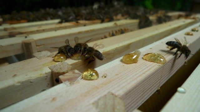
How badly the colony is affected as a result will depend on the percentage of bees infected – if a high percentage are infected then it can have a serious effect on the colony. The symptoms to watch out for are a colony that fails to build up in spring, with lower brood production than you would normally expect. Honey production is also affected, due to a decrease in foraging time spent as bees die off early. Colonies usually survive and have fewer infected individuals once warmer late spring weather appears and bees are able to fly out to excrete away from the hive.

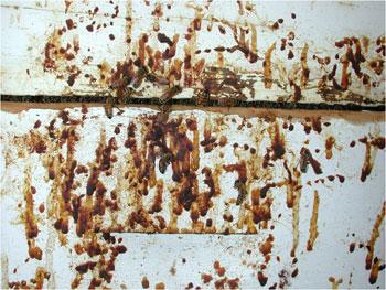
Some slight differences have been observed in the symptoms caused by the two species Nosema ceranae and Nosema apis. Dysentery spots and visible adult bee mortality in front of the hives are sometimes associated with N.apis but reported to be generally absent with N.ceranaeN.apis appears to thrive in colder weather whereas N.ceranae, which originated in Asia, seems to prefer warm climates and is more common during summer months. 

Could nosema ceranae be implicated in colony collapse disorder (CCD)?

There is some evidence that N.ceranae infections are more voracious than Nosema apis; in Spain N. ceranae infections have been observed to be characterised by a progressive reduction in the number of bees in a colony until the point of collapse. As the colony becomes under increasing pressure, secondary diseases frequently appear, including chalk brood and American foul brood.

There has even been some speculation that N. ceranae could be a contributing factor in CCD – if the colony breaks down, the queen can be found surrounded by a few bees, confusedly attending to brood that is already sealed. Infected bees tend to altruistically prevent spreading the infection by flying away and not returning to the hive. Newly emerged bees may not test positive for nosema, allowing the culprit to go undiscovered. However, more recent research has failed to confirm a link and researchers in the US have concluded that it is not the primary cause of CCD.

b) how you would confirm that the disease is present

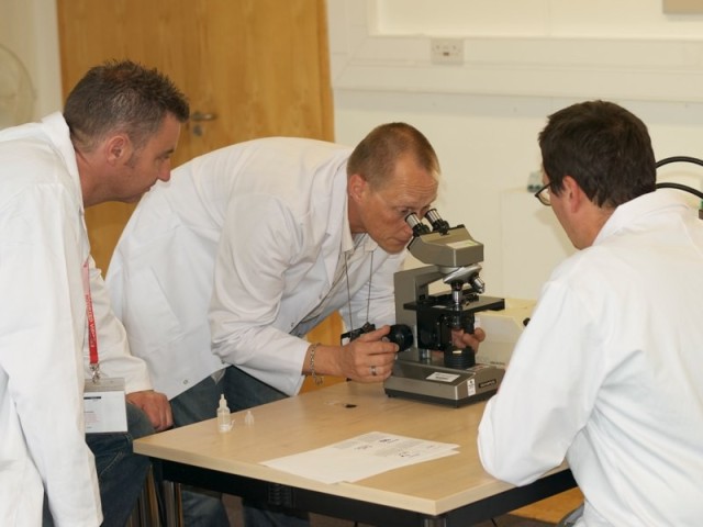
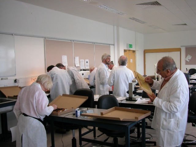
Definite confirmation requires microscopic examination of the abdomens of older adult bees, which can be collected from the hive entrance. Infection is highly variable in a colony, but foragers tend to contain the higher spore loads. Collecting bees leaving to go on cleansing flights rather than returning will be more effective. Send a sample of 30 bees to a microscopist or the UK National Bee Unit service in a small sturdy box (not plastic, which causes rapid sample degradation).

From a Beecraft magazine article – Bee Craft, Nosema ceranae Jan 2008:

“Abdomens of worker bees are ground up in a mortar with a little water and a drop of the resulting liquid spread onto a microscope slide. The tell-tale rice grain shapes can be seen using a microscope with a magnification of 400x. If you take a large enough sample of bees, you could probably detect minute levels of Nosema in many colonies. For a realistic result, it is recommended that you use around 30 bees.”

Diagram below from Scientificbeekeeping.com. The difference between the two species is very slight, but N.apis tends to have a more transparent centre and N.ceranae more of a slender, ‘bent’ shape.
nosema spores

The extent of infection is determined by counting the spores on a microscope grid and calculating the average number of spores per area and estimating from that the number of spores per bee.

Under the light microscope the spores of N. apis and N. ceranae appear as white/green, rice shaped bodies. Telling the difference between the two species is not easy, but some say an expert eye can spot it. On average N.apis spores are one micron larger in length than N.ceranae. Additionally N.ceranae spores can appear ‘bent’ and thinner than N.apis spores; however treatment is the same for both species. Genetic examination is the only reliable technique to distinguish between the two.

c) the action that should be taken to treat the disease if confirmed  

Fumadil B, the antibiotic previously used for treating nosema, has recently become no longer approved for sale in the UK (since December 2011). Without this treatment at our disposal, moving the colony onto fresh comb that contains no nosema spores is the best option.

Our best defence against nosema is good husbandry practices such as regularly changing brood comb, avoiding crushing bees through clumsy handling (this can release millions of nosema spores inside the hive) and maintaining strong well fed colonies, headed by young prolific queens.

As always, avoid moving brood frames between hives and do not feed honey from other colonies to bees – nosema spores can remain latent over a year on combs and for three to four months in honey. Wash hive tools between hives with a solution of one part washing soda to five parts water (e.g. 1kg of soda dissolved in 5 litres of water). Bee keepers should also consider re-queening susceptible colonies with queens from more tolerant stocks of bees better able to cope with Nosema infection.

d) how you would get the bees onto new sterile combs to allow you to remove the original frames for sterilisation  – give two methods.

Bailey comb exchange – all infected brood comb is removed gradually, over a period of about a month. A new brood box filled with foundation is placed over the existing one. Once the bees have drawn out some of the foundation, the queen is found and placed in the new brood box. A queen excluder is placed between the two brood boxes, so that she cannot move down to the old one. Heavy 2:1 mixture syrup should be fed to encourage the workers to draw out new comb quickly.

Three weeks later, once the larvae in the old brood combs have emerged, these frames can be removed and either burned or fumigated with acetic acid. Acetic acid is very corrosive stuff, so beginners should be cautious when trying this and seek advice from more experienced beekeepers first. The Bailey comb exchange is best done in early spring, such as March in the UK.

See the FERA Beebase website’s Replacing Comb factsheet for a more detailed explanation of the Bailey exchange method.

Shook-swarm – this is a dramatic spring clean for the bees, removing all their old brood comb with larvae cocoons/faeces, varroa and possibly nosema spores and giving them fresh new foundation to build on. Best done on a warm, sunny day in early Spring. 

First move your hive to one side and put a ‘new’ hive and foundation frames in the location of your old one, with a queen excluder on top of the floor (between the floor and the brood box). The queen excluder stops the queen absconding in the first week, and putting the new hive in the old location ensures that any foragers return to the right place.

Next, find the queen (easy, right?!) and put her safely in your pocket in a queen cage. Remove four frames from the centre of the new hive and place them to one side. Shake all the workers into the gap and return the queen. The old brood comb can now be burnt. Feed 2:1 strength sugar syrup to the colony to help them draw out new comb quickly and return regularly to top this up. The queen excluder can be removed a week later.

See the FERA Beebase website for a free downloadable shook-swarm factsheet which is worth following step-by-step if you’re shook-swarming for the first time.

B2. a) give a simple account of the structure and function of the alimentary, excretory and respiratory systems of the adult honeybee worker

  • Ailmentary systems

Digestion and excretion are the functions of the alimentary canal and its associated glands. Immediately inside the bee’s mouth the canal extends into a cavity with muscular attachments to the front of the head, so that it can expand and contract, providing small amounts of suction to help pass food from the proboscis and into the oesophagus. Waves of contractions are produced by muscles in the oesophagus, working nectar back into the bee’s initial ‘honey stomach’ or ‘crop’, where it is stored for a while.

At the end of the honey stomach is an important valve called the proventriculus, which stops the nectar going further until it’s needed.The proventriculus has four lips that are in constant movement, sieving out solids such as pollen grains and spores from the nectar and passing them back as a fairly dry lump into the ventriculus (mid-gut).

When the bee needs sugar, the proventriculus opens up and allows some nectar through into the ventriculus, where enzymes break the nectar down into molecules small enough to be passed through the gut wall into the haemolymph.Any waste residue is passed into the small intestine and held as faeces in the rectum until the bee is able to leave the hive for a poop. During long periods of cold weather in winter the rectum can expand to almost the whole length of the abdomen before the bee is able to fly out. This is when bees are more likely to have trouble ‘holding it in’, especially if they are suffering from dysentery.

Illustration below by R.E. Snodgrass from http://www.extension.org. Alimentary canal of worker (Phy-Rect), together with pharyngeal glands (1Gl), and salivary glands of head (2Gl) and of thorax (3Gl), as seen by cutting body open from above and pulling the ventriculus (Vent) out to left.

  • Excretory systems

As a bee digests its food, the food breaks down and the resulting products are circulated by the bee’s haemolymph (a bee’s equivalent to our blood). The useful products provide energy and bodybuilding substances. The waste products need to be expelled by the excretory system, which acts as a sophisticated filtering system. As well as removing waste substances, it also adjusts the levels of particular substances in the haemolymph, ensuring the correct balance between water and salts and that osmotic pressure and acidity remain within narrow limits.

  • Respiratory systems

The bee breathes through tubes called trachea, which carry oxygen throughout the bee’s body to where it’s required. In more complex animals such as mammals, oxygen is transported to body tissues by blood, but the haemolymph of insects is not involved in transporting oxygen.

The trachea open to the air around the bee through holes in the cuticle called spiracles, which in many cases are provided with a closing mechanism. Air enters the tracheal system through the spiracles and is diffused around the bee’s body.

Diagram below of the internal anatomy of a honey bee found at chdphd.com/PhD/Chapter2.php, adapted from Dade (1977) and Winston (1987).

honey bee anatomy diagram
b) explain how Acarine, Nosema and Amoeba affect these systems.

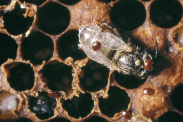
      • Acarine is a small mite named Acarapis woodi which lives in the main thoracic trachea (respiratory tubes) of the honey bee. A fertilised female will find her way into a bee’s trachea and lay 5-7 eggs soon after the bee emerges from its cell – she prefers her host to be only one to two days old. There is some evidence that she prefers drones as hosts, due to their larger tracheas.
Tracheal mite

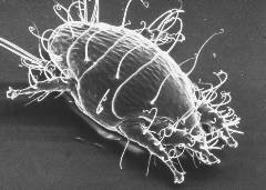
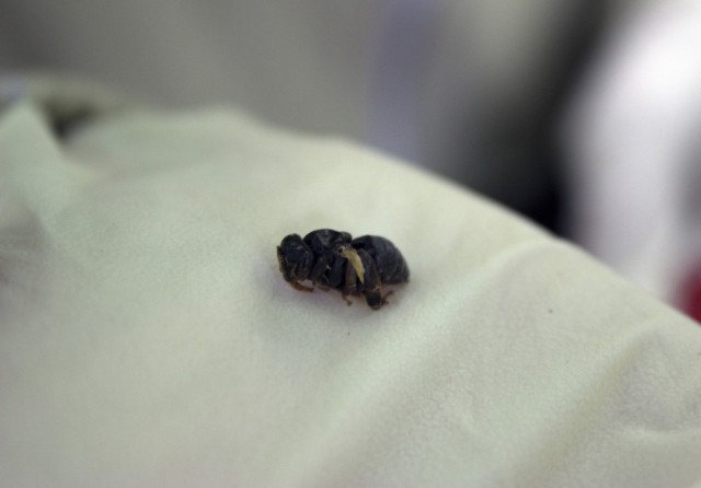
Tracheal mite/tiny guinea pig. Courtesy The Food and Environment Research Agency (Fera), Crown Copyright.

The eggs hatch in about five days, and the little larvae (which Ted Hooper describes in Guide to Bees and Honey as reminding him of “tiny guinea pigs”) develop into adult mites about nine days later. The mite family feed on the haemolymph (blood equivalent) of the infested bee through the walls of the trachea, piercing it with their mouth parts. Their life cycle is complete with fourteen to nineteen days, after which they mate in the trachea and the fertilised females leave to start a new family in other young bees.

Grabbing a new host

The female mites find a new host by leaving the trachea, clinging onto one of the bee’s hairs with one or two hind legs, and then waving their remaining legs around until a suitable young bee less than nine days old comes along. She will be able to tell by the smell of the bee and also because the trachea is protected by hairs which are not so dense in young bees. She grabs the hair of her new host with her front legs and is drawn into the first spiracle through wing vibrations and puffs of air. Guinea pigs never did anything so dastardly!

Effect on the colony

The poor bee can have a trachea stuffed full of the mites, which has the effect of blocking its trachea, shortening its lifespan. Some honey bee sub-species are more susceptible than others – acarine is a bigger problem in the US, where many beekeepers use Italian/New Zealand crosses. Bees native to the UK appear to have evolved a tolerance to the mite.

It is thought that acarine usually weakens a colony and makes it susceptible to other diseases rather than killing colonies outright. The National Bee Unit give a guideline figure for the U. K. that if 30% of bees in a colony are infested then the colony will die in the following spring, but say that generally in Europe a realistic threshold could be set at about 10%.

It was probably a viral infection such as Chronic Bee Paralysis Virus and not acarine (as was originally thought) that killed so many colonies in the infamous Isle of Wight disease outbreak of 1904.

Diagram below of tracheal mite life cycle found on the Mid Atlantic Apiculture Research Consortium website, @MAARECnews

tracheal mite lifecycle

  • As described above, Nosema spores multiply in the ventriculus (reaching 30 – 50 million spores) and impair the digestion of pollen, thereby shortening the life of the bee.

The spores germinate in an ingenious way – inside each spore is a coiled filament that is attached to one end of the spore. This filament everts quickly from the spore and injects a host mid-gut cell, pouring the contents of the spore (nucleus, membranes, etc.) into the host cell cytoplasm.

  • Amoeba is caused by a parasite named Malpighamoeba mellificae. Its cysts are ingested by nurse/cleaning bees and germinate in the rectum.

The spores migrate to the malpighian tubules (the bee’s ‘kidneys’), attack the cells lining the tubules and after 3-4 weeks will divide to create more cysts, that then accumulate in the rectum and are excreted. The cycle then begins again when hive bees ingest the cysts whilst cleaning.

So the cysts take longer to develop than nosema spores – and the number produced is also sixty times less. A bee also has numerous malpighian tubules, so not all the tubules are likely to be damaged.

Whether amoeba infection has much effect on the bee is not clearly known. However, infections are very often found in association with nosema and it is likely that a dual infection will be more damaging to the health of the honey bee. Considering how many really nasty parasites and pests honey bees have to fight, amoeba cysts appear to be pretty benign parasites. Hurray! Their cuddly round shapes are often seen under the microscope when examining a sample for nosema spores.

Microscope picture below from Scientificbeekeeping.com. Amoeba cysts are larger and more oval than nosema spores, and don’t glow in the center.

Printer-friendly pdf version.

References:

Posted in Disease prevention | Tagged , , | 11 Comments